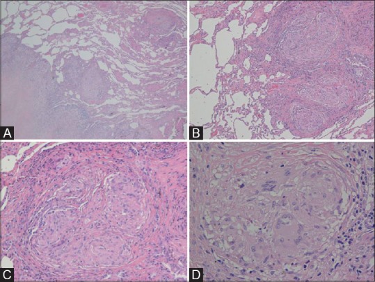
Figure 2(A-D).

The thoracoscopic biopsy shows an interstitial fibrotic lesion with a reticular and nodular pattern (A, H and E stain, magnification 40×) and a subpleural and septal distribution. The lesion is characterized by multiple non-confluent granulomas (B, 100×) accompanied by collagen deposition devoid of significant inflammation and necrosis (C, 200×). The granulomas is made of epitheliod hystiocytes with few multinuclear giant cells (D, 400×)
