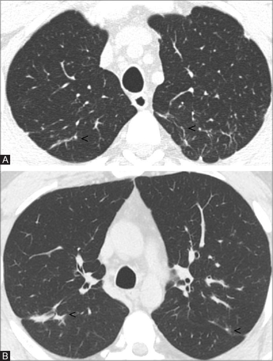
Figure 3(A and B).

Two years later follow-up HRCT images. Bilaterally, some small linear opacities persist (A and B; see arrowheads), no further parenchymal abnormalities are appreciable

Two years later follow-up HRCT images. Bilaterally, some small linear opacities persist (A and B; see arrowheads), no further parenchymal abnormalities are appreciable