
. 2017 Jul 7;2017:17-0057. doi: 10.1530/EDM-17-0057
 This work is licensed under a
 Figure 2.
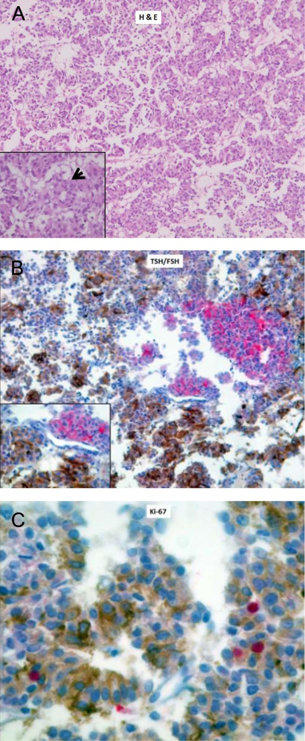
Histopathology and immunohistochemistry of the excised tumor. (A) H & E staining (40×, 100× in the inset) of the tumor showing a trabecular pattern and occasional pseudorosette formation (black arrow). (B) Double immunostaining with anti-TSH (brown) and anti-FSH (red) antibodies, labeled with diaminobenzidine and alkaline phosphatase, respectively, showing two distinct populations of cells (40×, 100× in the inset). (C) Ki-67 proliferative index (MIB-1) of 1–2% (positive nuclei are stained in red), mainly restricted to TSH-positive cells (400×).