
Figure 5. Physiologic levels of PD-L1 are not sufficient to prevent immune elimination of highly immunogenic unedited T9 sarcoma cells.
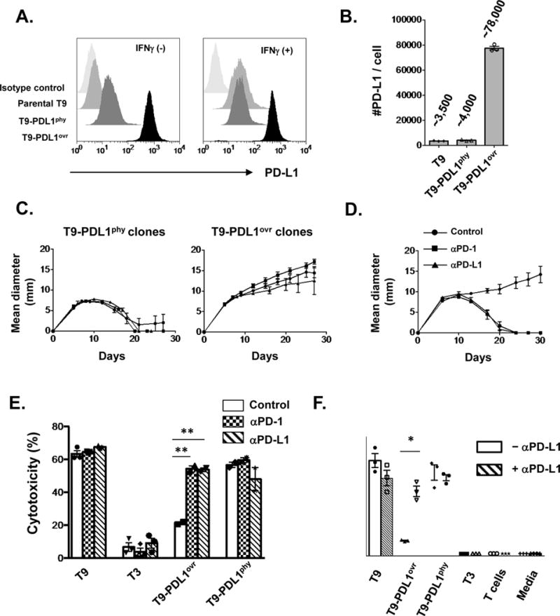
A) In vitro PD-L1 expression with or without IFNγ on T9 sarcoma cells constitutively expressing ectopic PD-L1 either at physiologic levels comparable to that induced by IFNγ on parental T9 cells (T9-PDL1phy) or those at high level over-expression (T9-PDL1ovr). Data are shown from at least three independent experiments. B) Numbers of PD-L1 molecules expressed on cells after IFNγ treatment in vitro. Data are shown by mean ± s.e.m of technical triplicates from at least three independent experiments. C) In vivo growth of three clones of T9-PDL1phy or T9-PDL1ovr cells in WT mice. Data are shown by mean ± s.e.m from at least two independent experiments (n = 5). D) Anti-PD-1 (4H2) or anti-PD-L1 (14D8) leads to tumor rejection in T9-PDL1ovr-bearing mice. Data are shown by mean ± s.e.m from two independent experiments (n = 5). E) In vitro cytotoxicity assay of mutant Spectrin-b2-specific CTL (C3) against tumor cells with anti-PD-1(4H2)/anti-PD-L1(14D8) blockade. Data are shown by mean ± s.e.m of technical triplicates (T9, T3) and duplicates (T9-PDL1ovr, T9-PDL1phy) from at least two independent experiments. F) In vitro IFNγ secretion from mutant Spectrin-b2-specific CTL (C3) against tumor cells with or without anti-PD-L1 (14D8). Data are shown by mean ± s.e.m of technical triplicates from at least two independent experiments. Samples in E, F were compared using an unpaired, two-tailed Student t test. **P < 0.01, *P < 0.05.