
Figure 2. Anatomical reorganization of CS connectivity for antagonistic muscle pairs during development.
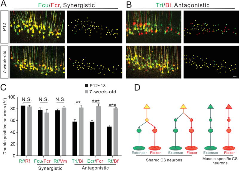
(A–B) Representative images (left: confocal and right: imaris reconstruction) of CS neurons labeled by dual-color PRV tracing from a pair of synergistic muscles (Fcu/Fcr, A) and a pair of antagonistic muscles (Tri/Bi, B) at P12 (top panels) and 7-week-old (bottom panels) wild-type mice.
(C) Percentage of double-labeled CS neurons in early postnatal (black bar) and adult mouse (grey bar) muscle pairs (synergistic and antagonistic) following dual-color PRV tracing. Significant increases in double-positive CS neurons were observed in adult animals for all antagonistic muscle pairs, but not synergistic muscle pairs or the Rf/Rf control injections: Rf/Rf, P18 (n=3), adult (n=6), P=0.7017; Fcu/Fcr, P12 (n=4), adult (n=4), P=0.5495; Rf/Vm, P18 (n=6), adult (n=4), P=0.2748; Tri/Bi, P12 (n=4), adult (n=5), P=0.0053; Ecr/Fcr, P12 (n=6), adult (n=6), P=0.0003; and Rf/Bf, P18 (n=6), adult (n=6), P<0.0001.
(D) Model summarizing the connectivity of disynaptic CS circuits. Where “n” represents the number of mice used in each experiment. Scale bar, 50 μm (B). See also Figure S2.