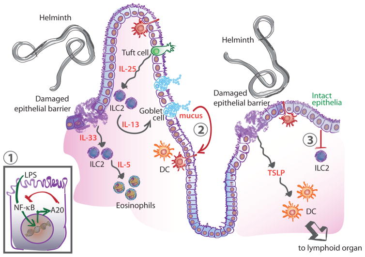
Figure 1. Regulation of Type 2 Immunity at Barrier Tissues.
Immune stimulus (e.g. helminth) leads to tissue damage and secretion of the cytokines IL-33 and TSLP. TSLP activates DCs to adopt a Th2-promoting phenotype and migrate to lymphoid organs to activate T cells. IL-33 signals on ILC2 leading to the recruitment of eosinophils through the secretion of IL-5. ILC2s also sense the increased production of IL-25 by tuft cells and secrete IL-13 which feeds back on goblet cells leading to increased mucus production. Negative regulatory mechanisms at the barrier include inhibition of NF-κB signaling in epithelial cells through the action of A20 (1). During homeostasis, mucus limits inflammation by inducing a regulatory pathway in DCs in the context of antigen uptake (2). Similarly, an intact epithelium provides immunoregulatory signals that limit ILC2 activation (3). Abbreviations: DC, dendritic cell; IL, interleukin; ILC2, type 2 innate lymphoid cell; TSLP, thymic stromal lymphopoietin.

