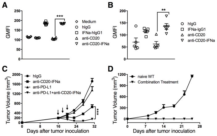
Figure 6. Anti-PD-L1 can enhance the therapeutic effect of anti-CD20-IFNα to control advanced B-cell lymphoma.
(A) A20 cells (5 × 10⁴) per well were incubated with 20 pM of indicated antibodies in 96 round-well plates (n = 6/group). 48 hours later, tumor cells were collected and the expression of PD-L1 was analyzed by flow cytometry. (B) WT BALB/c mice (n = 5/group) were injected subcutaneously (s.c.) with A20 cells (3 × 10⁶). A total of 3 μg of IFNα-IgG1, anti-CD20, anti-CD20-IFNα, or control hIgG was administered intratumorally (i.t.) on day 16. The average tumor size was 193 mm3. Two days later, tumor cells were collected and PD-L1 expression was analyzed by flow cytometry. (C) WT BALB/c mice (n = 13/group) were injected s.c. with A20 cells (3 × 10⁶) and treated i.t. with 3 μg of anti-CD20-IFNα or control hIgG on days 16, 18 and 20. 50 μg of anti-PD-L1 (10F.9G2) or Rat IgG (rIgG) was administered i.t. on the same time. (D) Approximately 45 days after the tumor rejection from (C), the mice were rechallenged with A20 cells (1.5 × 10⁷). Naïve WT BALB/c mice were used as the control. Data represent mean ± SEM of two independent experiments. **P < 0.01, ***P < 0.001.

