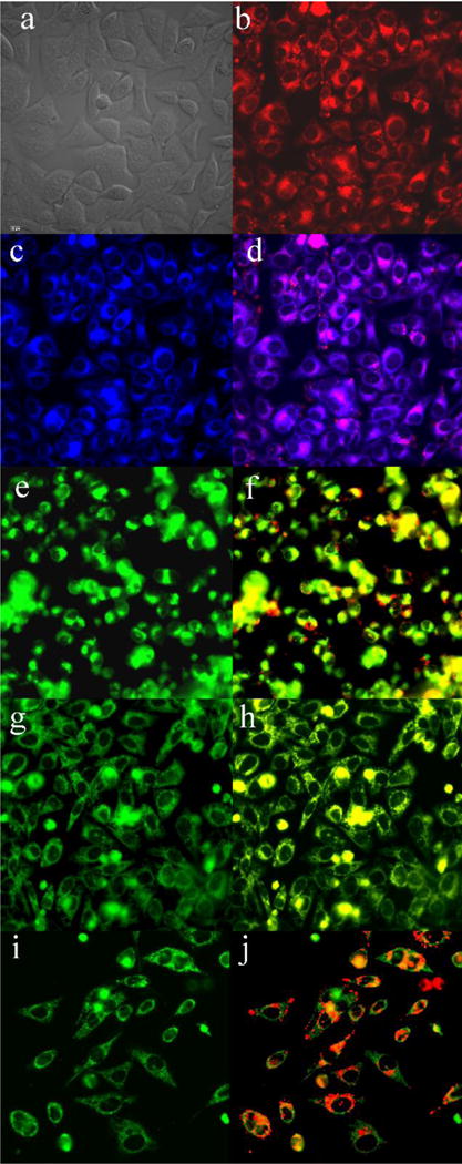
Figure 9.

Subcellular localization of 6b in HEp2 cells at 10 μM for 6 h. (a) phase contrast, (b) overlay of 6b and phase contrast, (c) ER Tracker Blue/White, (d) overlay of 6b and ER Tracker, (e) BODIPY Ceramide, (f) overlay of 6b and BODIPY Ceramide, (g) MitoTracker Green, (h) overlay of 6b and MitoTracker, (i) LysoSensor Green, (j) overlay of 6b and LysoSensor Green. Scale bar: 10 μm.
