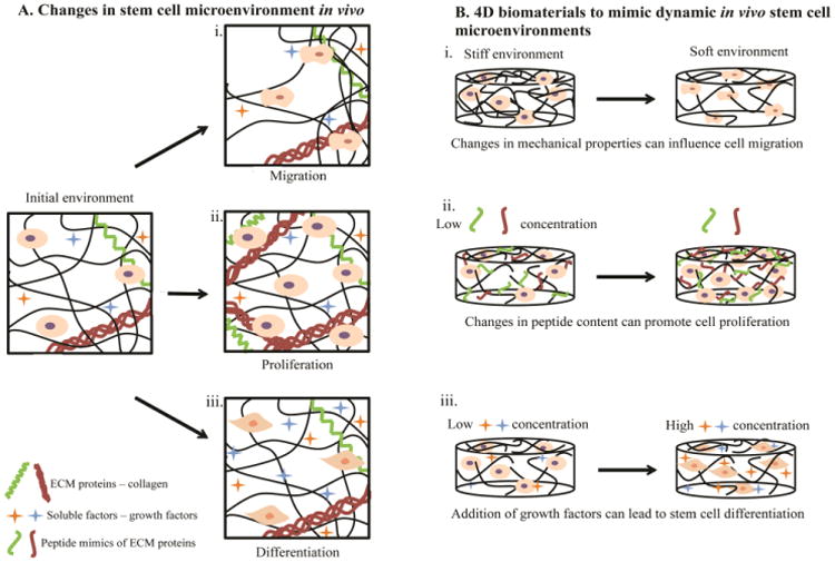
Figure 1. Changes in stem cell microenvironments in vivo captured by 4D biomaterials in vitro.

A) The microenvironment of stem cells in the body changes with time. These changes have been observed to modulate cellular fate and functions, such as i) migration in response to gradients of matrix density/stiffness, ii) proliferation in response to matrix remodeling, and iii) differentiation in response to soluble factors (e.g., growth factors). B) Creating materials that capture such changes aids in the study of how cells respond to microenvironment remodeling events, such as wound healing or disease progression, toward ultimately directing these processes. For example, 4D hydrogel-based biomaterials have been engineered to enable i) changes in the mechanical properties of synthetic matrices by the addition or removal of crosslinks, influencing cell migration throughout the materials. At higher crosslink densities and moduli, cells have been entrapped within hydrogel-based matrices (left), whereas at lower crosslink densities and moduli cell migration has been observed (right). ii) Variation in biochemical content within the hydrogels through addition or removal of biochemical moieties (e.g., integrin-binding peptides or protein fragments) has been observed to promote cell proliferation. iii) Addition or sequestration of growth factors swollen within or tethered to the synthetic matrix has been observed to regulate stem cell differentiation. Note, these examples are meant to be representative, rather than comprehensive, of the ways 4D biomaterials have and can be used to probe stem cell processes; many extracellular cues influence multiple cellular functions (e.g., growth factors can influence migration, proliferation, and differentiation).
