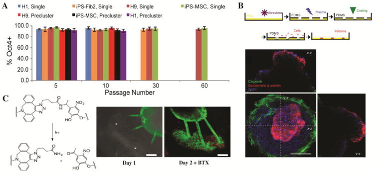
Figure 4. Stem and progenitor cell processes in 4D biomaterials.

A) Long term expansion of hPSCs has been achieved in thermoreversible PNIPPAAm-PEG hydrogels. Seven hPSC lines were cultured for multiple passages within these materials with ∼ 95% pluripotency, indicated by Oct4 expression. The longest hPSC line passage accumulated to ∼ 1072 -fold expansion at 60 passages or 280 days. Adapted from Lei and Schaffer with permission from Proceedings of the National Academy of Sciences USA PNAS [57]. B) 3D cardiac microchamber was formed by confinement of iPSCs within PEG microwells. Cell-laden PEG microwells (top) were fabricated by PEG polymerization, PDMS mask and etching of the PEG film, coating of wells, and seeding of cells. Cells (bottom, nuclei blue) in the center differentiated into cardiomyocytes, as indicated by sarcomeric α-actinin stain (red). Cells along the perimeter differentiated into myofibroblasts, as indicated by calponin stain (green). x-z and y-z cross section projections (above and to the left) show the inner void space, indicating a 3D cardiac microchamber. Adapted from Healy and coworkers with permission from Nature Communications Publishing Group [77]. C) EB ESMNs were co-encapsulated with C2C12 cells within a photodegradable hydrogel. Channels (10 μm ×10 μm) were degraded between these two cell types using a 740-nm two-photon laser to cleave the o-nitrobenzyl-containing crosslinker (left). At two days, synapses (stained with alpha-bungarotoxin, red) were observed between motor axon extensions (green) and myotubes (right). Adapted from Anseth and coworkers with permission from Biomacromolecules the American Chemical SocietyACS Publications [37].
