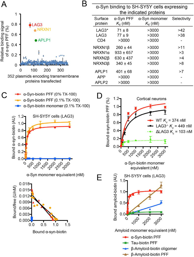
Fig. 1. α-Syn PFF binds to LAG3.
(A) Individual clones from a library consisting of 352 individual cDNAs encoding transmembrane proteins (GFC-transfection array panel, Origene) were transfected into SH-SY5Y cells, and the relative binding signals of human α-syn PFF to individual transmembrane proteins are shown. Positive candidates are LAG3 (NM_002286), NRNX1 (NM_138735) and APLP1 (NM_005166). (B) Mouse α-syn-biotin monomer and α-syn-biotin PFF binding affinity to SH-SY5Y cells expressing the indicated proteins. LAG3* Kd assessment was performed without Triton X-100. All other experiments were performed with 0.1% Triton X-100. Transmembrane proteins similar to the candidates were also tested. Quantification of bound α-syn-biotin PFF to the candidates was performed with ImageJ. Kd values are means ± SEM and are based on monomer equivalent concentrations. Selectivity was calculated by dividing Kd (monomer) by Kd (PFF). Binding of α-syn-biotin monomer was detected at a concentration of 3000 nM, but binding was not saturable. (C) α-Syn-biotin monomer or α-syn-biotin PFF binding to LAG3-overexpressing SH-SY5Y cells as a function of total α-syn concentration in 0% Triton X-100 (TX-100) or 0.1% TX-100 conditions (monomer equivalent for PFF preparations, top panel). Scatchard analysis (bottom panel). Kd= 71 nM (0% TX-100) and 77 nM (0.1% TX-100), data are the means ± SEM, n = 3. (D) Binding of α-syn-biotin PFF to cultured cortical neurons (21 days in vitro (DIV)) is reduced by LAG3 knockout (LAG3−/−), as assessed by alkaline phosphatase assay. α-Syn-biotin PFF WT-Kd = 374 nM, LAG3−/−-Kd = 449 nM, estimated Kd for neuronal LAG3 [dashed line: ΔLAG3 = wild-type (WT) minus LAG3−/−] is 103 nM. Data are the means ± SEM, n = 3. * P < 0.05, Student’s t-test. Power (1-β err prob) = 1. (E) Specificity of LAG3 binding with α-syn-biotin PFF (Fig. S4). Tau-biotin PFF (Fig. S8), β-amyloid-biotin oligomer and β-amyloid-biotin PFF (Fig. S9) are negative controls.

