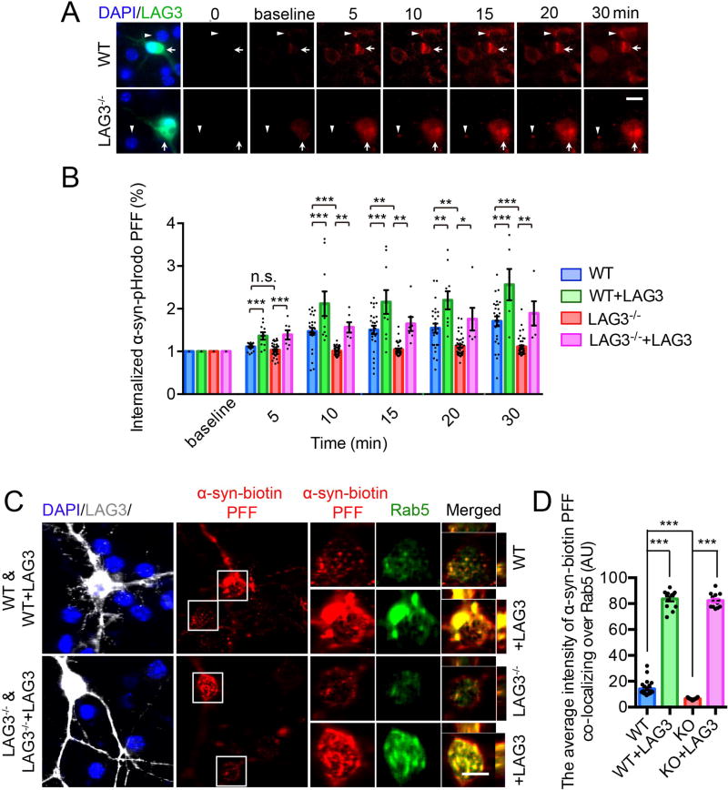
Fig. 2. Endocytosis of α-syn PFF is dependent on LAG3.
(A) Live image analysis of the endocytosis of α-syn-pHrodo PFF. α-Syn PFF was conjugated with a pH dependent dye (pHrodo red), in which fluorescence increases as pH decreases from neutral to acidic environments. White triangles indicate non-transfected wild-type (WT) or LAG3−/− neurons and white arrows indicate LAG3 transfected neurons. Scale bar, 10 µm. (B) Quantification of panel A, cell number (5–46) from n = 3. (C) Internalized α-syn-biotin PFF co-localizes with Rab5. Co-localization of internalized α-syn-biotin PFF and Rab5 was assessed by confocal microscopy, scale bar, 10 µm. (D) Quantification of panel C, cell number (13–32) from n = 4. One-way ANOVA with Tukey’s correction. Data in B and D are as means ± SEM. *P < 0.05, **P < 0.01, ***P < 0.001. Power (1-β err prob) = 1.

