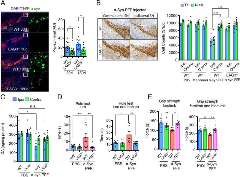
Fig. 6. α-Syn PFF induced pathology is reduced by deletion of LAG3 in vivo.
(A) Representative P-α-syn immunostaining and quantification in the substantia nigra par compacta (SNpc) of WT and LAG3−/− mice sacrificed at 30 and 180 days after intrastriatal α-syn PFF injection. Data are the means ± SEM, n = 5–9 mice per group, one-way ANOVA with Sidak’s correction. (B) Stereology counts from TH immunostaining and Nissl staining of SNpc DA neurons of WT and LAG3−/− mice at 180 days after intrastriatal α-syn PFF, α-syn monomer or PBS injection. Data are the mean number of cells per region ± SEM, n = 5–9 mice per group, one-way ANOVA with Dunnett’s correction. (C) DA concentrations in the striatum of α-syn PFF-injected mice and PBS-treated controls measured at 180 days by HPLC. Data are the means ± SEM, n = 5–8 mice per group, one-way ANOVA with Tukey’s correction. (D, E) 180 days after α-syn PFF injection, the pole test and grip strength was performed in WT or LAG3−/− mice injected with PBS or α-syn PFF. Behavioral abnormalities in the pole test and grip strength induced by α-syn PFF injection were ameliorated in LAG3−/− mice. Data are the means ± SEM, n = 7–9 mice per group for behavioral studies. Statistical significance was determined using one-way ANOVA with Tukey’s correction, * P < 0.05, ** P < 0.001, *** P < 0.001, n.s., nonsignificant. Power (1-β err prob) = 1.

