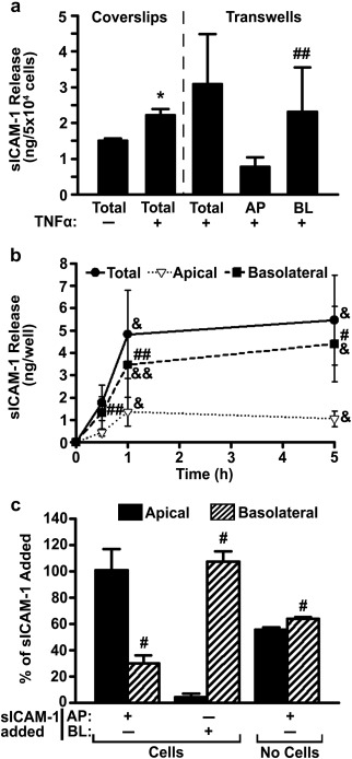
Figure 1.

Release of sICAM‐1 by ECs. (a) HUVECs were grown on coverslips or transwell inserts in control medium versus medium containing TNFα (16 h). Cells were then washed and sICAM‐1 release into the cell medium [apical (AP), basolateral (BL), and total (AP + BL)] was examined after 30 min, using ELISA. (b) Distribution of sICAM‐1 release by TNFα‐activated HUVECs grown on transwells was similarly measured at 30 min, 1 h, or 5 h. (c) Relative distribution of exogenous sICAM‐1, 4.5 h after its addition to the AP or BL chambers of transwells in the absence of cells versus the presence of TNFα‐activated HUVECs. Data are mean ± SEM. *Comparison to non‐activated ECs; #comparison between apical and basolateral chambers at each time point; &comparison to 30 min (one symbol is p < 0.1 by Student's t‐test and two symbols is p < 0.1 by Mann‐Whitney Rank Sum test)
