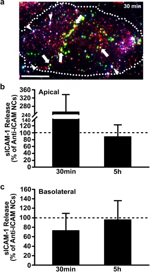
Figure 6.

Inhibition of anti‐ICAM NC endocytosis attenuates sICAM‐1 release. (a) Image of TNFα‐activated HUVECs grown on coverslips and incubated for 30 min at 37°C with green fluorescent anti‐ICAM NCs. Nonbound NCs were removed by washing, and then the cells were fixed and immunostained (see Materials and Methods) to render surface‐bound NCs triple labeled in green + blue + red (white color; arrowheads). Instead, internalized membrane ICAM‐1 complexed with NCs appears double labeled in green + red (yellow color; arrows) and internalized NCs without membrane ICAM‐1 are labeled in green alone. Scale bar = 10 µm. (b) Apical and (c) basolateral release of sICAM‐1 by TNFα‐activated HUVECs grown on transwell inserts and incubated with anti‐ICAM NCs in the presence of amiloride, an inhibitor of CAM endocytosis. Incubations were for 30 min (pulse), followed by anti‐ICAM NC removal and incubation for additional time up to 5 h (chase). Data are expressed relative to absence of amiloride (control; horizontal dashed line). Data are mean ± SEM
