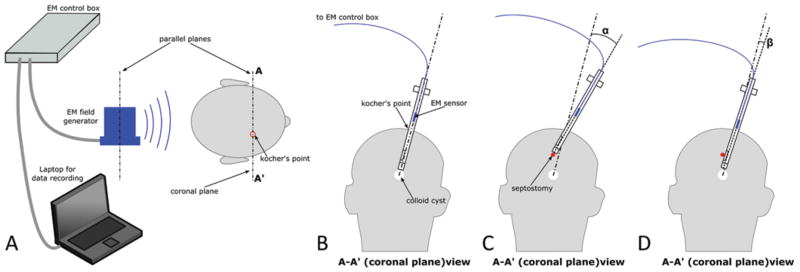
FIG. 6.
Measurement of brain deformation during ventricular neuroendoscopy. Setup of the electromagnetic (EM) tracker (A), insertion path for the colloid cyst resection (B), instrument pivoting (measured by angle α) required for septostomy using a standard rigid scope (C), and pivoting (measured by angle β) required using a multiport endoscope (D).

