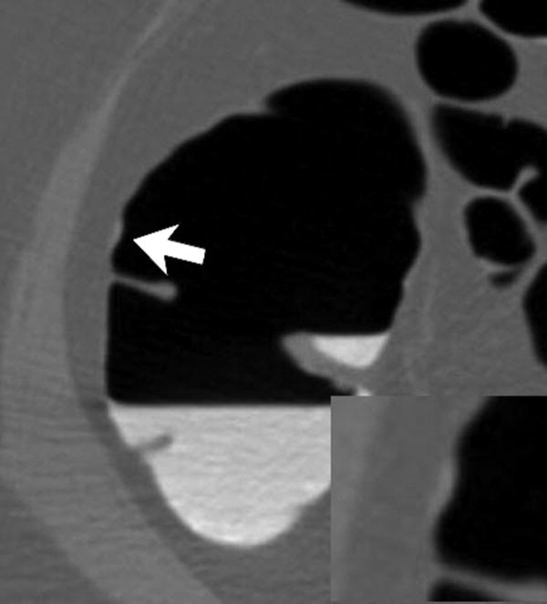
Figure 2.
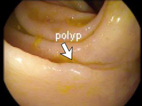
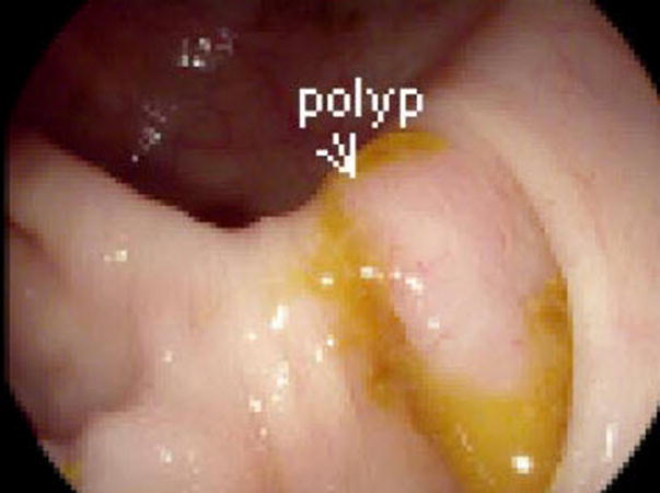
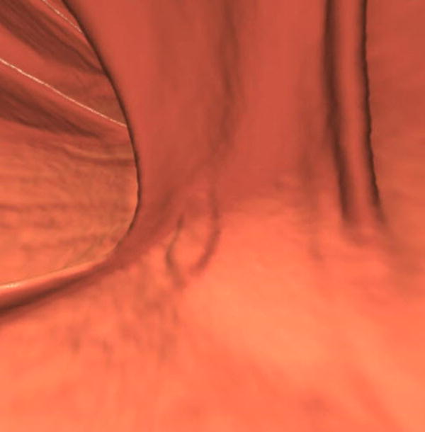
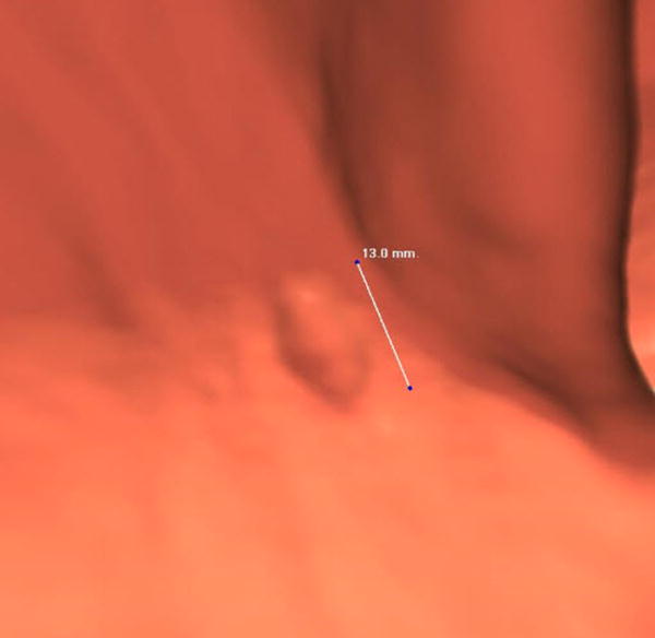
Three different patients underwent screening CTC and had a right-sided SSA. Each patient has a corresponding optical colonoscopy image (A, D, G), 3D CTC endoluminal image (B, E, H), and a 2D CTC axial image (C, F, I). The optical colonoscopy images nicely depict the flat, subtle nature of these large right-sided polyps. The 3D CTC endoluminal images show the appearance at imaging that led to the polypectomy at colonoscopy. Notice that the polyps are more prominent and protruding, as compared with the colonoscopic appearance. Axial 2D CTC images demonstrate how the 3D appearance represents a combination of a flat polyp and the overlying adherent tagging contrast agent (arrows). Magnified images (insets in F and I) better depict the subtle soft tissue thickening underneath the contrast agent.

