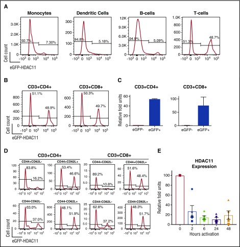
Figure 1.
HDAC11 expression is downregulated in activated cells and Teff. (A) Utilizing EGFP-HDAC11 reporter mice, EGFP expression was assessed by flow cytometry in various immune cells. (B) Expression of EGFP-HDAC11 was further assessed in CD4+ (left panel) and CD8+ (right panel) T cells obtained from the lymph nodes. Plots shown are representative of 3 mice assessed in 3 independent experiments. (C) EGFP expressing and nonexpression cells were flow sorted and analyzed by qRT-PCR for expression of HDAC11 mRNA. Error bars are from 3 technical replicates per group. (D) Expression of EGFP was evaluated by flow cytometry in CD4+ and CD8+ T-cell subsets defined by CD44 and CD62L expression. Plots shown are representative of 3 mice assessed in 3 independent experiments. (E) WT CD3+ T cells isolated from mouse lymph nodes were left unstimulated or activated by αCD3/CD28 beads for indicated times and assessed by qRT-PCR for HDAC11 mRNA expression. Combined results from 5 mice, each represented with a unique symbol, assessed over 2 independent experiments are shown. P values of baseline vs activation comparisons are as follows: 2 hours: P < .05; 6 hours: P < .001; 24 hours: P < .01; 48 hours: P < .05.

