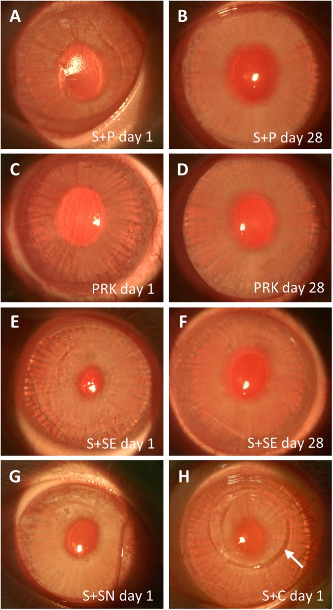
Fig 1. Slit lamp photographs of cornea captured after SMILE retreatment.
(A) Rough surface was noted on the cornea on day 1 after PRK (S+P day 1). (B) Cornea appeared clear with smooth surface on day 28 after PRK (S+P day 28). The appearance of the corneas was comparable to those in the PRK day 1 (C) and PRK day 28 (D) groups, respectively. (E) Cornea appeared clear, but the surface became rougher on day 1 after the secondary SMILE (S+SE day 1). (F) The minor surface undulation was not seen 28 days after secondary SMILE (S+SE day 28). (G) Cornea appeared clear and the surface was smooth after secondary SMILE when the lenticule was not extracted (S+SN day 1). (H) Cornea appeared clear after flap creation by Circle pattern and subsequent stromal excimer ablation (S+C day 1). Flap side cut created by the Circle pattern, which extended beyond the small incision (arrow), was visible.

