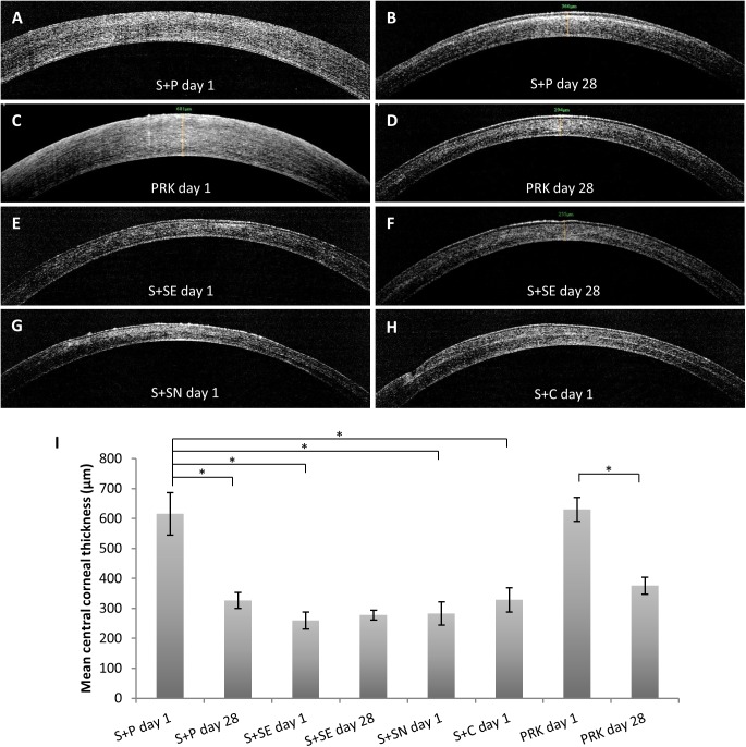
Fig 2. Cross-sectional visualization of cornea captured after SMILE retreatment.
(A) Cornea appeared edematous on day 1 after PRK (S+P day 1). (B) Sub-epithelial stromal haze was observed on day 28 after PRK, although the cornea appeared thinner (S+P day 28). (C) Reciprocal observation in S+P day 1 could be made on cornea treated with PRK only on day 1. (D) In contrast to S+P day 28 group, stromal haze was not seen in PRK day 28 group. Corneal cap interface was hardly visible after secondary SMILE either in the groups which had the lenticule extracted (both S+SE day 1 and S+SE day 28 groups) (E and F) or the group with lenticule left not extracted (S+SN day 1 group) (G). (H) Corneal flap interface was visible as a reflective plane on day 1 after refractive enhancement utilizing VisuMax Circle software (S+C day 1 group). (I) Bar graph showing mean central corneal thickness after each retreatment method. Resulting corneal thickness was analyzed using one way ANOVA and a post hoc Tukey comparison procedure. *p<0.001.

