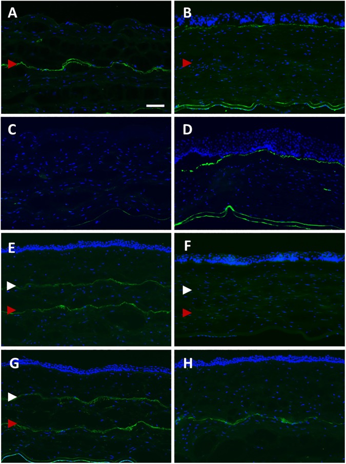
Fig 5. Expression of fibronectin in the central cornea after SMILE retreatment.
(A) Fibronectin was not present at the superficial layer of the exposed stroma on day 1 after enhancement with PRK (S+P day 1 group), but appeared relatively strong along the primary SMILE interface located in the posterior stroma (red arrowhead). (B) On day 28, fibronectin was localized in the sub-epithelial layer of the cornea (S+P day 28 group). (C) Fibronectin staining was not present in any layer of the stroma on day 1 after PRK only treatment (PRK day 1 group). (D) Similar to S+P day 28, the staining was localized in the sub-epithelium in PRK day 28 group. (E) The interface resulted from the primary SMILE (red arrowhead) and from the secondary SMILE (white arrowhead) expressed fibronectin (S+SE day 1 group). (F) Post-secondary SMILE day 28, no expression of fibronectin was observed in the stroma (S+SE day 28 group). (G) In S+SN day 1 group, both primary SMILE interface and secondary SMILE cap cut expressed fibronectin. (H) Staining was seen along the flap interface in S+C day 1 group. Scale bar = 50 μm.

