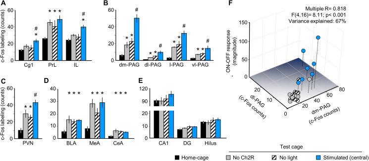
Fig 2. MRR stimulation selectively activates brain areas involved in emotional control.
Optic stimulation selectively increased the expression of the activity marker c-Fos in two sub-regions of the medial prefrontal cortex (A), the whole periaqueductal gray (B), and the paraventricular nucleus of the hypothalamus (C). PrL and the amygdala was activated by cage transfer, but not stimulation (D); the hippocampus showed no responses (E). Panel F is a 3D illustration of the Multiple Regression analysis presented in the text. BLA: basolateral amygdala; CA1: CA1 region of the hippocampus; CeA: central amygdala; Cg1: anterior cingulate cortex; DG: dentate girus of the hippocampus; dl-: dorsolateral; dm-: dorsomedial; IL: infralimbic cortex, l-: lateral; MeA: medial amygdala; PAG: periaqueductal gray; PrL: prelimbic cortex; PVN: paraventricular nucleus of the hypothalamus; vl-: ventrolateral. * p<0.05 significant difference from home-cage controls; # p<0.05 significant difference from “no ChR2”.

