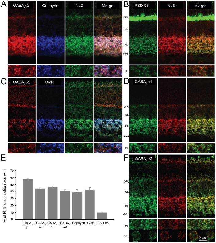
Fig 2. Localization of NL3 in the mouse retina.
To ascertain the distribution of NL3 at retinal synapses, co-immunolabelings with excitatory (B) and inhibitory (A, C, D, F) postsynaptic markers were carried out. NL3 essentially did not associate with the excitatory postsynaptic protein PSD-95 (B, E); it colocalized extensively with the ubiquitous GABAAγ2 receptor marker (A, E) and equally well with GABAAα1, α2 and α3 receptor subsets, suggesting its association with diverse retinal GABAA receptor subtypes (C-F). NL3 was also frequently observed together with glycine receptors (GlyR, labeled with a pan-GlyR antibody) (C, E). Plots in E represent true colocalization estimates after subtraction of random associations (see Methods). N = 3 animals and at least 4 image sections analyzed per staining. Plots represent mean ± SEM. OPL, outer plexiform layer; INL, inner nuclear layer; IPL, inner plexiform layer; GCL, ganglion cell layer.

