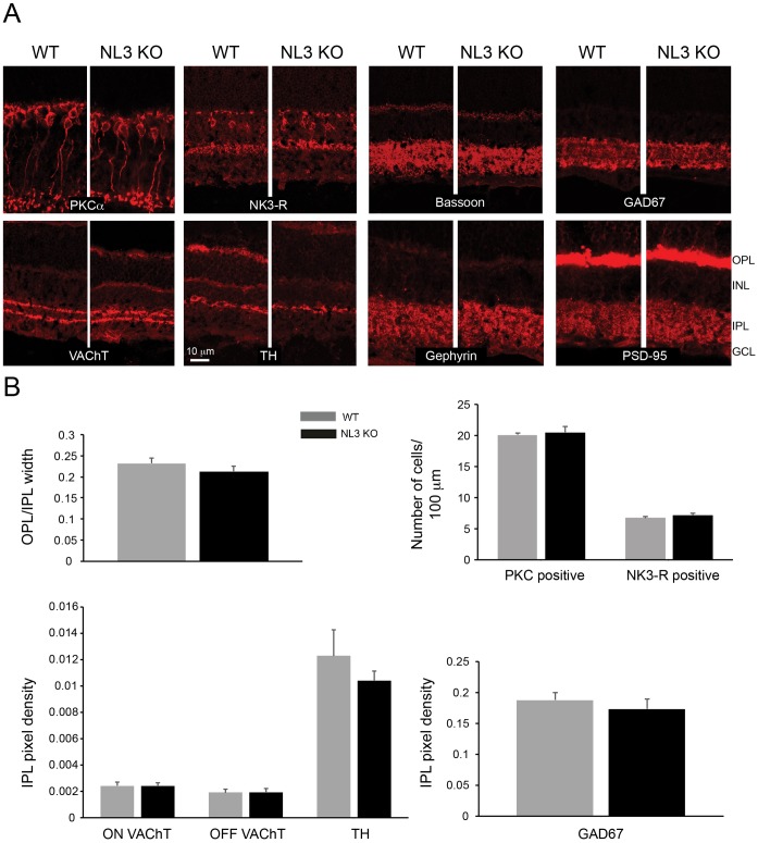
Fig 3. Architecture of the NL3 KO retina.
Rod (PKCα) and OFF-cone (NK3-R) bipolar cells, cholinergic (VAChT), dopaminergic (TH) and GABAergic (GAD67) amacrine cells are similarly represented and organized in WT and NL3 KO retinae (A). The overall synaptic connectivity is intact in the absence of NL3, as illustrated by a comparable layout of ribbon synapses at the OPL and conventional synapses at the IPL (bassoon), and excitatory (PSD-95) and inhibitory (gephyrin) postsynapses in WT vs. NL3 KO retinae. Moreover, the relative thickness of the synaptic plexiform layers, as well as the number of PKC and NK3-R positive cells and the density of labeling for VAChT (both in the ON and OFF sublamina), TH and GAD67 were comparable across genotypes (N = 3 WT-KO littermate pairs, at least 5 images per sample) and point towards an intact retinal architecture of the NL3 KO (B). OPL, outer plexiform layer; INL, inner nuclear layer; IPL, inner plexiform layer; GCL, ganglion cell layer.

