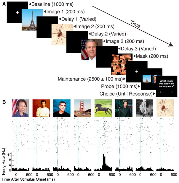
Figure 1. Experimental Design and Example Response.
(A) Behavioral task. In each trial, the subject saw a stream of four or three images, chosen from a pool of eight or nine, respectively, for 200 ms each. In three-quarters of trials, a blank screen was presented after each image, such that the inter-stimulus interval was either 200, 500, or 800 ms. In the remaining one-quarter of trials, there was no intervening blank screen. Following image presentations, subjects saw a mask followed by a fixation cross, which was presented for a minimum of 2.4 s. The fixation cross then disappeared, and subjects saw two probe pictures simultaneously, one of which had been presented in the preceding stream of images. After these probes disappeared, the subject pressed a key to signal the previously presented image.
(B) Spike rasters and peri-stimulus time histograms for a stimulus-selective single unit recorded from the right entorhinal cortex at sample presentation, including presentations at all inter-stimulus intervals. Images at the top indicate presented stimuli.

