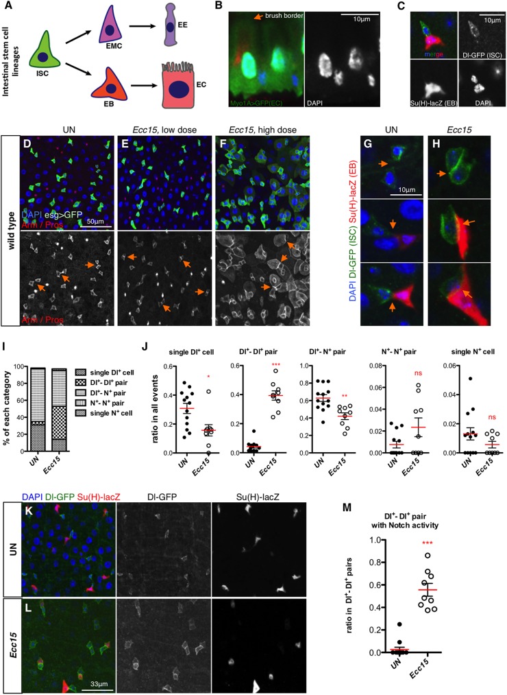
Fig 1. Increased progenitor contact is a general feature of regenerating intestine.
(A) Current model of intestinal stem cell (ISC) lineages. Cell types in the Drosophila midgut include ISC, enterocyte (EC) and its precursor enteroblast (EB), enteroendocrine cell (EE) and its precursor enteroendocrine mother cell (EMC). (B) Basal-apical (bottom to top) organization of the Drosophila midgut epithelium. ECs are visualized by Myo1A>GFP (Green) and brush border is indicated. (C) A typical ISC-EB pair in wild type midgut of an unchallenged fly. ISC is detected with Dl-GFP (green) and EB with Su(H)-lacZ (red). (D-F) Representative intestines of unchallenged (D) flies, flies orally infected with a low dose (E) or a high dose of Ecc15 (F). (G-H) Representative progenitor pairs in unchallenged control (G) and Ecc15-infected (H) intestines. Junctions between progenitor pairs are indicated with orange arrows in (D-H). (I-J) Summary of all the progenitor combinations in unchallenged control (n = 969, from 13 guts) and Ecc15-infected (n = 654, from 9 guts) intestines, and ratios of each category (including single Dl-GFP+ cell, Dl-GFP+—Dl-GFP+ pair, Dl-GFP+—Notch+ pair, Notch+—Notch+ pair and single Notch+ cell, from left to right) in all the events under both conditions. (K-L) Representative images of unchallenged control (K) and Ecc15-infected (L) intestines. Note that the increase in progenitor contact and presence of many Dl-GFP+—Dl-GFP+ pairs with one cell showing weak Notch activity are detected in (L). (M) Ratio of Dl-GFP+—Dl-GFP+ pairs with Notch activity (n = 42 in control and 259 in Ecc15 challenged guts). Each dot represents one gut.

