. 2017 Jun 20;6:e25328. doi: 10.7554/eLife.25328

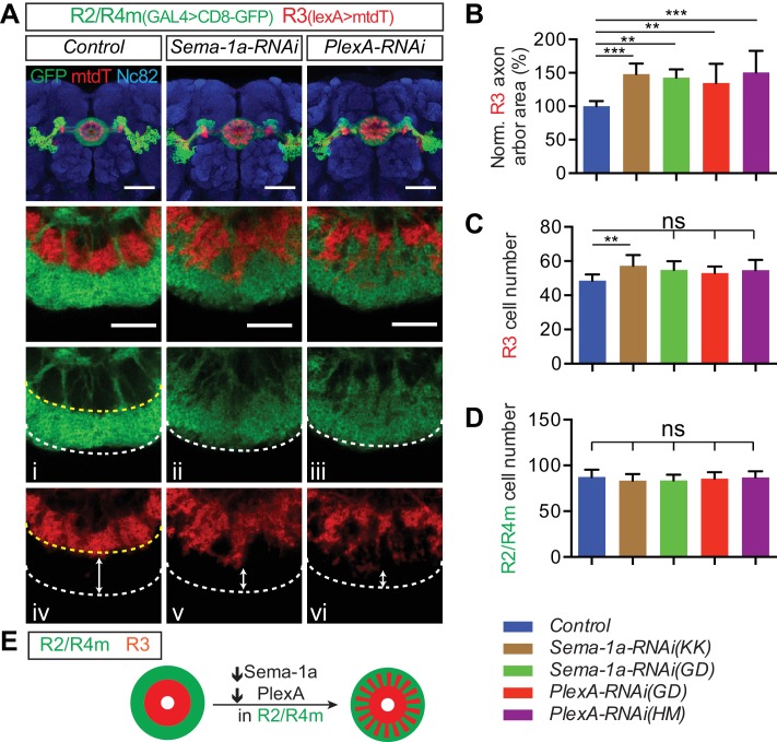
Figure 5. Sema-1a and PlexA are required in R2/R4m neurons to constrain R axon growth within the ellipsoid body.

(A–D) R2/R4m neurons were labeled by R32H08-GAL4-driving CD8-GFP, while R3 axons were labeled using R54B05-lexA-driving mtdTomato (mtdT) in adult fly brains. Knocking down Sema-1a or PlexA in R2/R4m neurons did not change axon trajectory or cell numbers of either R3 or R2/R4m (panels C and D), but resulted in non-cell autonomous R3 axon arbor expansion in the EB (panel B). Seven to eight brains for each genotype were used for quantification. ‘ns’ p>0.1234; *p<0.0332; **p<0.0021; ***p<0.0002. See Figure 5—source data 1 for detailed statistical analyses. (E) Schematics showing that both R3 (red) and R2/R4m (green) axons expand when Sema-1a or PlexA is down-regulated in R2/R4m, leading to intermingled R3 and R2/R4m axons. Scale bars are 50 μm in low-magnification images in top panels and 20 μm in high-magnification images in other panels.

DOI: http://dx.doi.org/10.7554/eLife.25328.015

Figure 5—source data 1. Statistical analysis of R3 axon quantification in R2/4m RNAi.
DOI: 10.7554/eLife.25328.016
Figure 5—source data 2. Statistical analysis of R3 RNAi quantification.
DOI: 10.7554/eLife.25328.017
Figure 5—source data 3. Statistical analysis of single R3 MARCM quantification.
DOI: 10.7554/eLife.25328.018

Figure 5.

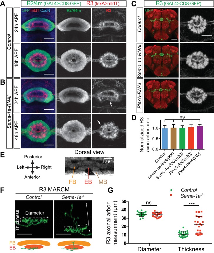
Figure 5—figure supplement 1. R2/R4m Sema-1a, but not R3 Sema-1a/PlexA, is required for R axon lamination.

Figure 5—figure supplement 1.

(A–B) R2/R4m and R3 axons were differentially labeled by R32H08-GAL4-driving CD8-GFP (green) and R54B05-lexA-driving mtdT (red), respectively. Knocking down Sema-1a in R2/R4m neurons led to gradual changes in R axon elaboration during early pupal stages. (C–D) R46D01-GAL4 was used to express CD8-GFP (green) and UAS-RNAi in a group of R3 neurons. Knockdown of either Sema-1a or PlexA did not change R3 axon elaborations. The R3 axonal arbor area was measured and compared to controls in panel D. Eight to ten animals were used for quantification of each genotype. All p-values are > 0.21. Detailed statistical analyses are available in Figure 5—source data 2. (E) The mushroom body (MB), the ellipsoid body (EB) and the fan-shaped body (FB) are adjacent to one another along the anterior-posterior axis in the adult Drosophila brain, shown here using Nc82 immunostaining. (F–G) R84H09-GAL4 was used to express CD8-GFP in R3 neurons in MARCM clones. In Sema-1a-/- single-cell MARCM clones, the radial elaboration of R3 axons within the EB is generally normal as indicated by no change of R3 axon ‘Diameter’ compared to control clones (34.83 ± 2.46 μm for the control (n = 24 SSCs) and 34.49 ± 2.53 μm for the Sema-1a-/- (n = 21 SSCs), p=0.826). However, some Sema-1a-/- R3 neurons extended a few axon branches posteriorly into the FB, which was quantified by assessing the increase in axon arbor ‘Thickness’ as observed in dorsal view images (10.83 ± 3.17 μm for the control (n = 24 SSCs) and 22.51 ± 9.78 μm for the Sema-1a-/- (n = 21 SSCs), p<0.001). See Figure 5—source data 3 for details of statistical analyses. Scale bars are 20 μm in panels A, B and F, and in right high-magnification images in panel C (black and white panels); 50 μm in left low-magnification images (color panels) in panel C.
Figure 5—figure supplement 2. Sema-1a and PlexA are not required for pb-eb-gall dendrite elaboration and synaptogenesis in the EB.

Figure 5—figure supplement 2.

(A) R2/R4m and pb-eb-gall neurons were differentially labeled using R32H08-GAL4-driving CD8-GFP (green) and R19G02-lexA-driving mtdT (red), respectively. Knocking down Sema-1a or PlexA in R2/R4m neurons led to changes in R2/R4m axon lamination, but no changes in pb-eb-gall dendrite elaboration in the EB. (B) R32H08-GAL4 and R19G02-lexA were used to express nSyb-spGFP1-10 and CD4-spGFP11 in R2/R4m and pb-eb-gall neurons, respectively. Sema-1a-RNAi was co-expressed in R2/R4m neurons in experimental conditions. And mtdT was co-expressed to labeled pb-eb-gall projections in all animals. (C–D) pb-eb-gall neurons were labeled by R19G02-GAL4-driving CD8-GFP (green). R2/R4m (red in C) and R3/R4d (red in D) neurons were labeled by R32H08-lexA and R70B04-lexA, respectively, driving mtdT. Knocking down Sema-1a or PlexA in pb-eb-gall neurons alters pb-eb-gall axon projections, but does not change pb-eb-gall dendrite elaboration and R axon lamination in the EB. Scale bars are 50 μm in low-magnification images in all panels; 20 μm in high-magnification images in panel A (i–vi) and B (i–iv).