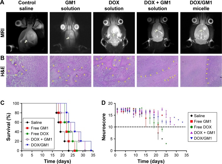
Figure 5.
MRI images (A), H&E staining (B, ×100), Kaplan–Meier plot (C), and neuroscore (D) of tumor-bearing rats after treatment with saline, free GM1 solution, free DOX solution, DOX + GM1 solution, and DOX/GM1 micelle, respectively. White arrows in MRI images indicate the tumor, and yellow arrows in H&E staining indicate tumor necrosis.
Abbreviations: DOX, doxorubicin; H&E, hematoxylin and eosin.

