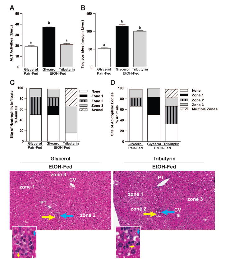
Figure 6. Effects of tributyrin on hepatic injury and zonation of neutrophilic infiltrate and acidophilic bodies following chronic-binge ethanol exposure.
Mice were treated as described in Figure 1. Mice were euthanized 9-hours post-ethanol gavage. A & B) Plasma was separated from blood collected from the posterior vena cava. Activity of ALT was measured in plasma. Hepatic triglyceride content was measured in whole liver homogenates. C–E) Liver was excised, fixed in formalin, embedded in paraffin and later stained with hematoxylin and eosin for histologic analysis. Quantitative assessment of the neutrophilic infiltrate and acidophilic bodies are shown. Bars represent percentage of mice in each group exhibiting the designated number of foci of neutrophilic infiltrates per high power field, acidophilic bodies and their zonal distribution in the liver (zone 1, 2 or 3; azonal: no recognizable pattern; multiple zones, multifocal but possible throughout the tissue with inconsistent distribution). Representative H&E histological sections of livers are shown for the study groups. Yellow arrows indicate neutrophilic infiltrates; blue arrows indicate acidophil bodies. (H&E stain, 100×).

