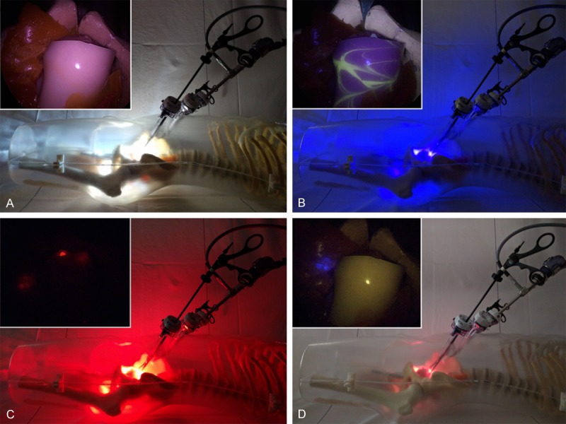
Figure 5.

Experimental set-up in a prostate cancer phantom. The outside view of the light source and laparoscopic positioning are shown with the laparoscopic image inserted: (A) White light setting for anatomical context, (B) Autofluorescence setting for the detection of fluorescein (nerves; yellow), (C) Cy5 setting for the detection of Cy5 (tumor lesions; red), and (D) ICG setting for the detection of ICG (sentinel nodes; blue).
