Figure 3.

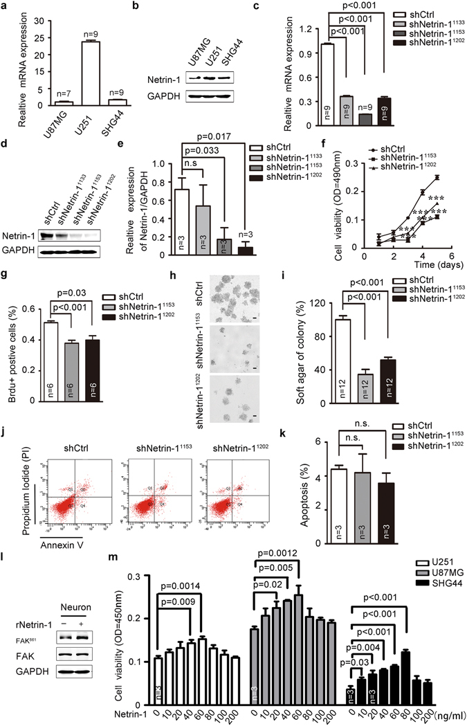
Netrin-1 was required for cell proliferation in cultured glioma cells. (a and b) Netrin-1 mRNA and protein levels were determined by real-time PCR and western blotting in three cultured glioma cells. (c and d) Detection of netrin-1 expression by real-time PCR and western blotting in U251 cells infected with lentivirus expressing shCtrl or shNetrin-1. GAPDH was used as a loading control in both assays. (e) Quantification of netrin-1 expression using Gel-Pro Analyzer 4 software. (f) MTT assays were used to determine the cell viability of U251 cells infected with lentivirus expressing shCtrl or shNetrin-1. (g) BrdU analysis was performed to detect cell proliferation in U251 cells infected with lentivirus expressing shCtrl or shNetrin-1. (h) U251-shCtrl or shNetrin-1 cells were plated in 0.3% soft agar. Three thousand cells were incubated for 16 days, and the media was changed every 2 days. Cells were counted using a dissecting microscope. Scale bar: 50 μm. (i) The colony formation assays were quantified by counting the number of colonies. (j and k) Apoptosis was measured by flow cytometry of U251 cells after infection with lentivirus expressing shCtrl or shNetrin-1. (l) Cells were treated with recombinant netrin-1. Phosphorylated FAK861 was detected by western blotting. (m) U251, SHG44 and U87MG cells were treated with increasing concentrations of recombinant human netrin-1. Cell viability was determined by CCK-8 assay after the addition of netrin-1 for 48 h. The data are shown as the mean ± SEM. n.s., P > 0.05.
