Figure 2.

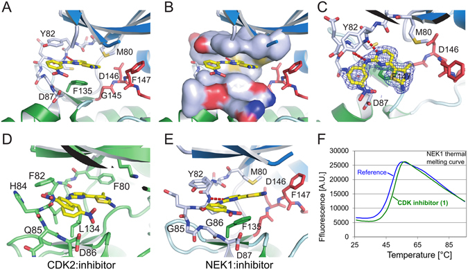
Inhibitor Binding to NEK1 kinase domain (A) Binding of CDK2/CDK9 inhibitor 1 to NEK1. The inhibitor is shown in yellow, and the DFG motif of NEK1 is shown in red. (B) As A, with a molecular surface shown around the NEK1 ATP binding site residues above and below the bound inhibitor, illustrating the tight fit of the inhibitor between Phe135, Met80 and Tyr82. (C) As A, viewed from above the inhibitor with an experimental 2Fo-Fc electron density shown contoured around the inhibitor at σ = 1.0. (D,E) CDK2 (PDB ID 1PXO) and NEK1 structures bound to inhibitor 1 and viewed from the same angle, showing the difference in binding angle of 1. (F) The inhibitor 1 was identified by its thermal stabilisation (ΔTm) of NEK1 kinase domain.
