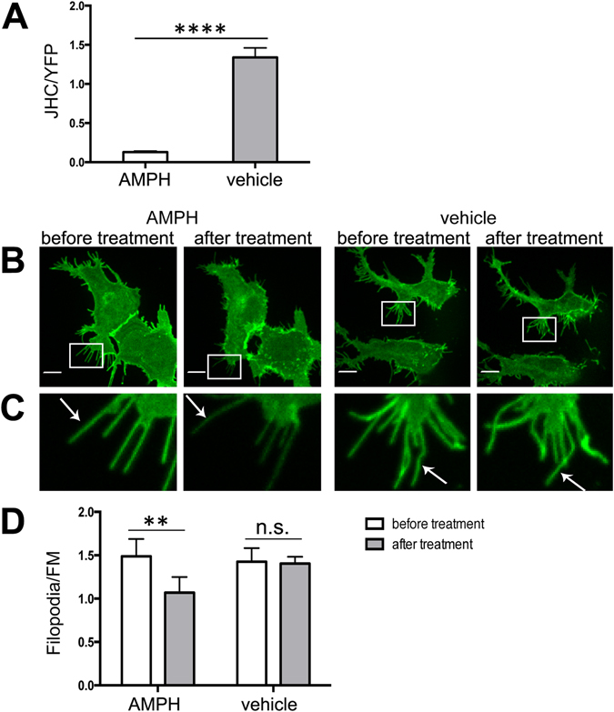
Figure 8.

AMPH treatment reduces the concentration of wt DAT in the filopodia of HEK293 cells. (A) Cells transiently expressing YFP-HA-DAT were treated with AMPH or vehicle (control) at 37°C for 1 hr. Cells were then incubated with 100 nM JHC1-64 for 10 min. Images were acquired through 515 nm (YFP, green) and 561 nm (JHC1-64, red) filter channels. The ratio of JHC1-64 and YFP fluorescence intensities (JHC/YFP) was calculated for individual cells. Results are shown as mean ± SEM, n = 5. ****p < 0.0001. (B) Cells transiently expressing YFP-HA-DAT were incubated with 10 μM AMPH or vehicle in KRH at 37 °C for 30 min. Live-cell imaging was performed through 515 nm (YFP, green) filter channel. Maximal z-projections of 5 consecutive x-y-confocal planes are shown. Scale bars, 10 μm. (C) Insets represent high magnification images of the regions marked by the white rectangle in (B). Arrows point on examples of peripheral filopodia. (D) The filopodia/FM ratios of YFP fluorescence intensities were calculated in experiments exemplified in (B,C). Results are shown as mean ± SEM, n = 8. **p < 0.01.
