
Figure 6.
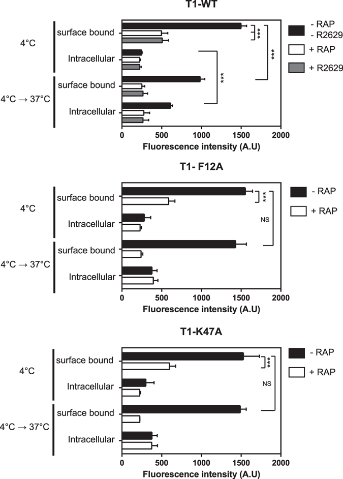
Biochemical analysis of LRP-1-mediated endocytosis of wild-type TIMP-1 and mutants. Cortical neurons from mouse embryos cultured for 48 h on poly-L-lysine-coated coverslips were incubated at 4 °C for 1 hour with 5 nM fluoT1-WT, fluoT1-F12A or fluoT1K47A in the presence or absence of 500 nM RAP or 30 µg/mL of blocking LRP-1 polyclonal antibodies (R2629). After careful washing, part of the cells was used to quantify the surface bound and intracellular signal at 4 °C. The other part was then incubated at 37 °C for an additional 10 min to quantify the surface bound and intracellular signal. Fluorescence intensity was quantified by spectrophotometry and expressed as arbitrary units (A.U.). Values below 300 A.U. are considered to be nonspecific. Error bars indicate mean ± SD, *** indicates significantly different with a P-value < 0.001 and NS indicates a value that is not statistically significant.