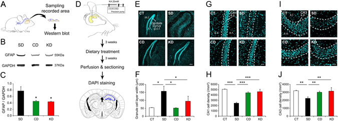
Figure 5.

CD treatment rescues cytological and molecular correlates of chronic epilepsy. (A) Tissue obtained from the recording site of KA mice was collected for immunoblotting. (B,C) Expression of GFAP was significantly reduced in mice treated with KD and CD (n = 4 per group, all p < 0.05). (D) KA mice were prepared for histological analysis of hippocampal area CA1, CA3 and DG. This article was published in The mouse brain in stereotaxic coordinates, Paxinos, G & Franklin, K, p.98, Copyright Elsevier Academic Press, 2001. This figure is not covered by the CC BY licence. Elsevier. All rights reserved, used with permission. (E,F) Both CD and KD (CD, n = 5; KD, n = 5) fed mice showed protection against dispersion of the granule cell layer (g.c.l.) compared to SD fed mice (SD, n = 6) and mice that did not receive KA (CT, n = 3).*p < 0.05. (G–J) CD and KD fed mice were also protected against cell loss in CA1 and CA3 compared to SD animals. **p < 0.01, ***p < 0.001. (h) hilus, m.l.: molecular layer, s.l.: stratum lucidum, s.o.: stratum oriens, s.p.: stratum pyramidale, s.r.: stratum radiatum.
