Figure 1.

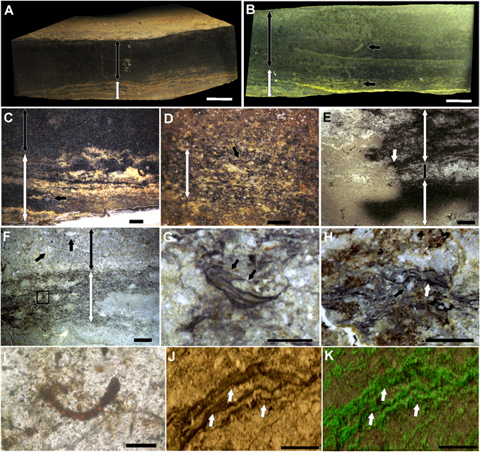
Biolaminites associated with Cloudina. (A) Polished hand sample showing the layer with microbial mat textures (white double-headed arrow) and the dark mudstone (black double-headed arrow). (B) Polished hand sample with a microbial mat layer (white double-headed arrow) and a dark mudstone layer (black double-headed arrow) and Cloudina oriented vertically (top black arrow) or inside microbial mats (bottom black arrow). (C) Polished hand sample with oxidized microbial mat textures (white double-headed arrow) associated with a Cloudina tube in cross-section (black arrow) and a mudstone layer above (black double-headed arrow). (D) Polished hand sample with microbial mat textures (white double-headed arrow) showing dark crenulated laminations (black arrow). (E) Thin section of microbial mat textures (white double-headed arrows) with an oxidation front (white arrow) and interbedded mudstones (black double-headed arrow). (F) Thin section demonstrating the presence of Cloudina inside crenulated laminations (black square and white double-headed arrow) at the base and associated with Cloudina (top black arrows) in a mudstone layer (black double-headed arrow). (G) Magnification of the black square in (F), showing a small individual of Cloudina inside the laminations and showing internal tubular structures (white arrow). (H) Thin section of the microbial mat textures showing dark lamination (white arrow) overlying occasional quartz grains (black arrow). (I) Microfossil of cyanobacteria. (J) Dark crenulated laminations (white arrows) of the microbialite associated with Cloudina. (K) Raman mapping of the region shown in (J) showing the presence of the “G” band of kerogen (green) mainly in the laminations (white arrows). Scale bars (A,B) 1 cm, (C,E,F) 2 mm, (D, G,H) 1 mm, (I) 20 µm (J,K) 0.2 mm.
