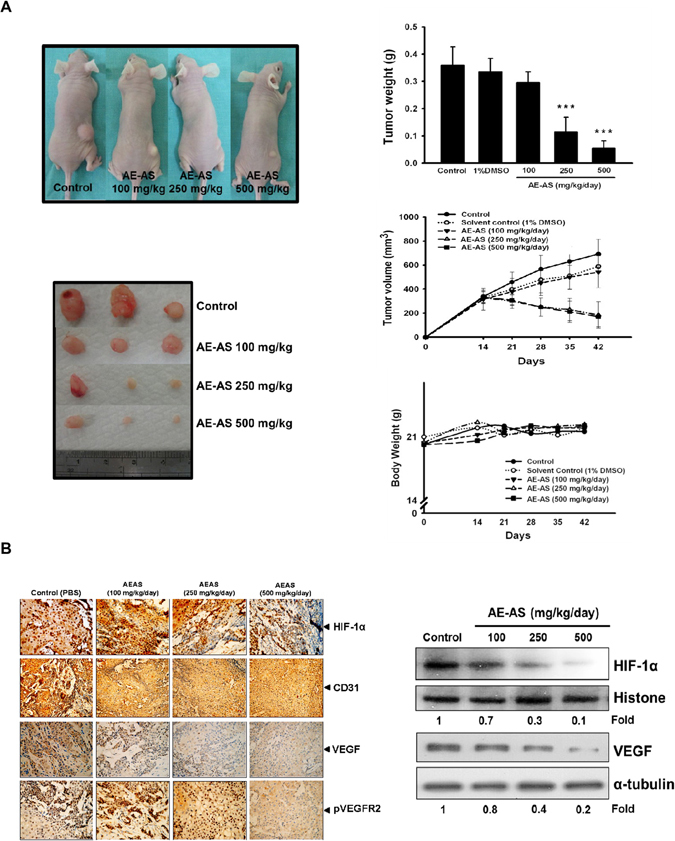
Figure 6.

AE-AS reduced tumor angiogenesis and growth. The mice were injected with T24 cells (s.c.) for 15 days followed by treatment with different doses of AE-AS for 30 days, the images of tumor sections, tumor size and weight, and body weight of mice were measured (A). The protein expression of HIF-1α, VEGF, CD31 and pVEGFR2 in tumor tissues was determined by immunohistochemical staining or Western blotting assay (B). Data was expressed as mean ± SEM (n = 5). ***P < 0.001 versus untreated cancer mice (Control group).
