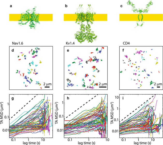
Figure 1.

Single-molecule trajectories and their TA MSD. (a) Structure of cockroach NavPaS protein (PDB accession code 5X0 M). This is the only solved structure for any eukaryotic Nav channel. (b) Structure of Kv1.2 assembled with the beta2 subunit (code 3LUT). (c) Structure of CD4 dimer (assembled from codes 1WIO and 2KLU). Structures were produced using PyMOL. The yellow bands in panels a–c represent the plasma membrane with the intracellular protein domains being below the membrane. (d–f) Trajectories in a representative cells obtained by single-particle tracking. (g–i) TA MSDs of the trajectories in panels (d–f). The dashed lines are guides to linear behaviour.
