
Figure 7. Knockdown of egfr-3 reduces asymmetric cell division during neoblast repopulation after sublethal irradiation.
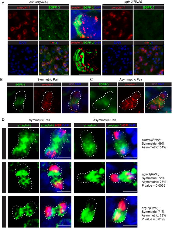
(A) Immunostaining with an antibody against EGFR-3 in control(RNAi) planarians. Egfr-3 knockdown eliminates the staining. (B) Representative images showing EGFR-3 and smedwi-1 distribution in symmetric cell division. (C) Representative images showing EGFR-3 and smedwi-1 distribution in asymmetric cell division. (D) Representative images and numbers of asymmetric vs. symmetric cell divisions in control(RNAi), egfr-3(RNAi), and nrg-7(RNAi) planarians at 14 dpi. Scale bar= 10 μm. p value calculated for egfr-3(RNAi) vs. control(RNAi) and nrg-7(RNAi) vs. control(RNAi), respectively, using Two tailed Fisher's exact test. See also Figures S7.