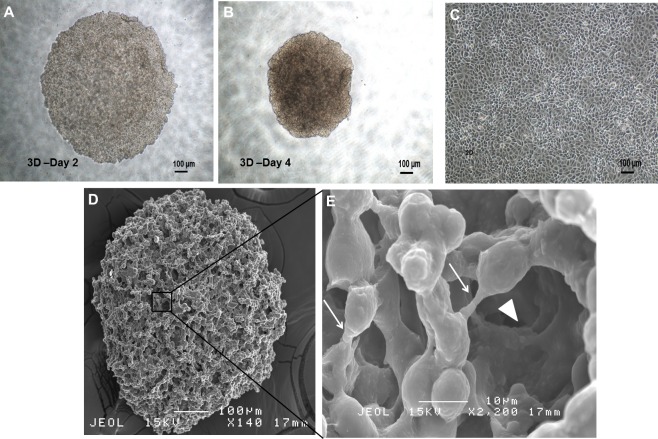
Figure 1. Spheroid and monolayer cells from estrogen-independent human breast adenocarcinoma cell line MDA-MB-231.
(A, B) Spheroid cells organised themselves into a compact and rigid three-dimensional structure 96 h post-culture in serum-free environment (magnification: 4×, scale bar; 100 µm). (C) Parental cells cultured in two-dimensional monolayer condition displayed as spindle shaped cells and adherent in nature (magnification: 4×, scale bar; 100 µm). (D) Ultrastructure analysis of spheroid cell coincided with clumping of cells to one another, with preservation of the overall structure of the spheroidal architecture (magnification: 140×, scale bar; 100 µm). (E) Higher SEM magnification showed the presence of cell–cell junctions (arrows) within the spheroid that are responsible for maintaining strong cell–cell contact (magnification: 2,200×, scale bar; 10 µm). Micropores indicated by ‘arrowheads’ allowed the exchange of nutrients across the inner layer of the spheroid with their surroundings.

