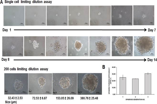
Figure 2. Secondary MDA-MB-231 spheroids formed using limited dilution techniques at single cell seeding and 200 cells/well.
(A) Generation of secondary spheroids from a single cell acquired microscopically for 14 days (magnification: 20×, scale bar; 100 µm). Secondary spheroids showed increased size spheroids from day 3 to day 14 (magnification: 20×, scale bar; 100 µm). Size of the spheroids and images are representative of three biological replicates. (B) Sphere-forming assay of secondary spheroids of MDA-MB-231 cell lines. SFE was counted from first to third generation. Data are based on the mean percentages of the number of spheres formed within a culture relative to the initial cell seeding number (means ± SD, n = 3)

