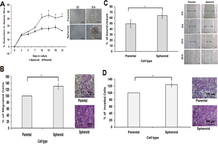
Figure 4. Spheroid cells demonstrated increased cell proliferation, migration, wound healing ability and invasion capacity relative to parental cells.
(A) Comparison of cell proliferation ability of the spheroid cells monitored using Alamar Blue assay. Determined by the reduction of Alamar Blue, spheroid cells showed significantly higher cell proliferation than dissociated parental cells when cultured in the same seeding number and condition. (B, C and D) Bar charts show higher cell migration, wound healing and invasion abilities in spheroid cells compared to their parental controls. Cell migration and invasion assays were measured by extracting the resulted stained cells and the absorbance of the dye colours were quantified. On the other hand, in wound healing assay, a gap was created and the cells’ proliferative ability to close the gaps were measured and recorded. The wound healing process of the spheroid and parental cells were taken at 0, 6, 12 and 24 h post-wound initiation.

