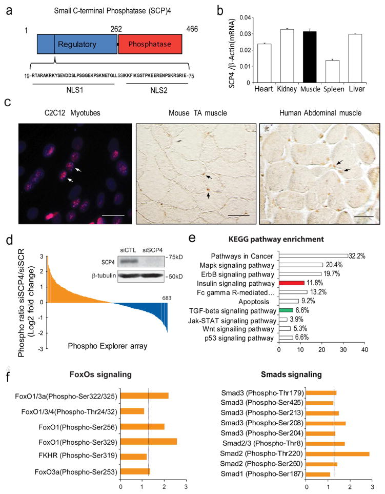
Figure 1. SCP4 may regulate FoxOs and Smads signaling in skeletal muscle cells.
a: Structural diagram of small c-terminal phosphatase 4 (SCP4); note that there are two nuclear location signaling (NLS) in its regulatory domain.
b: SCP4 mRNA expression in different tissues in mice (mean±SEM, n = 3).
c: Immunostaining revealed that SCP4 is mainly located in the nuclei of C2C12 myotubes (immunoflorescent staining, red: SCP4, blue: nuclear, scale bars: 20 μm) and myofibers from mice and human (DAB staining, brown: SCP4, Differential Interference Contrast (DIC) microscopy). Scale bars: 50 μm.
d: Phosphoproteome array in C2C12 myotubes with knockdown of SCP4. Data showing the fold change of induced phosphoproteins (yellow) and suppressed phosphoproteins (blue). The inserted immunoblots show siRNA knockdown efficiency.
e: KEGG pathway analysis of induced phosphoprotein genes (fold change > 1.5) indicated that insulin (in red) and TGF-β ( in green) signaling pathways were influenced by SCP4 knockdown.
f: The fold-change of phosphorylation sites in transcription factors FoxOs and Smads after SCP4 knockdown in C2C12 myotubes.

