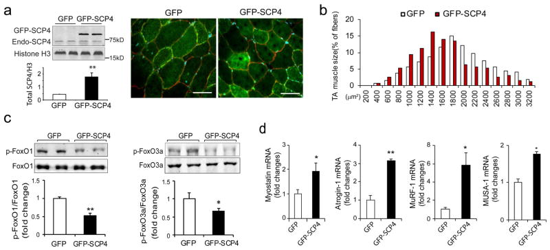
Figure 5. Forced expression of SCP4 in muscle results in myofiber atrophy in mice.
a: TA muscles of normal C57B6J mice were transfected with GFP control (GFP) and SCP4-GFP plasmids using electroporation. After 2 weeks, the total nuclear SCP4 (endogenous SCP4 plus SCP4-GFP) was assessed using immunoblots (n=5, **p<0.01); cryosections (6 μm) were immunostained with anti-dystrophin antibody to outline myofibers (red fluorescence). Scale bars: 50 μm.
b: The distribution of myofiber sizes in TA muscles transfected with SCP4-GFP was shifted leftward compared with the result in TA muscles electroporated with CTL-GFP. Data were obtained from 5 animals in each group.
c: Overexpression of SCP4 in TA muscles resulted in a decrease in p-FoxO1 and p-FoxO3a. A statistical analysis of immunoblots was shown on the right (mean±SEM, n = 5/group, **p<0.01,
d: The mRNA level of Myostatin, Atrogin-1, MuRF1 and MUSA1 were examined using RT-PCR in in TA muscles expressed GFP-CTL or GFP-SCP4 (mean±SEM, n = 5/group, **p < 0.01, *p<0.05)

