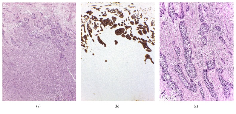
Figure 2.
Leiomyosarcoma and squamous cell carcinoma. (a) Squamous cell carcinoma at the upper part of leiomyosarcoma. Note an abrupt demarcation between two components (H&E, 4x). (b) Squamous cell carcinoma was highlighted by positive staining for AE1/AE3, whereas leiomyosarcoma was completely negative (IHC stain, 4x). (c) Infiltrative nests of squamous cell carcinoma surrounded by desmoplastic stromal reaction (H&E, 10x).

