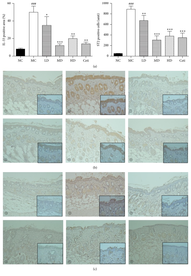
Figure 7.
(a) Results of immunohistochemical detection of IL-33 positive area and ST2 positive cells (×200). High magnification photo is in the black square frame (×400). Statistical results were expressed as mean ± SEMs, ###P < 0.001 compared with the normal group; ∗P < 0.05, ∗∗P < 0.01, and ∗∗∗P < 0.001 compared with the model group. (b) Detection of the expression of IL-33 protein in mouse dorsal skin tissue by immunohistochemistry. (c) Detection of the expression of ST2 protein in mouse dorsal skin tissue by immunohistochemistry. (①): normal control group; (②): model control group; (③): low-dose QRQS group; (④): middle-dose QRQS group; (⑤): high-dose QRQS group; (⑥): cetirizine medicine group.

