
Fig. 3.
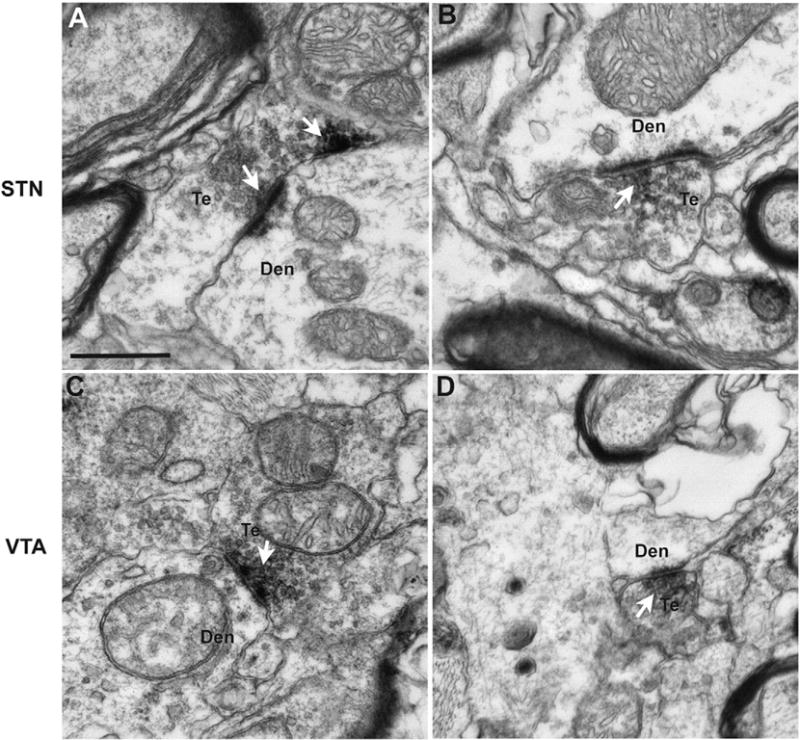
Electron micrographs of DβH-positive terminals (Te) that form asymmetric axo-dendritic synapses in the STN (A, B) and VTA (C, D) of a normal rhesus monkey. The arrows indicate aggregates of peroxidase deposit confined to pre-synaptic vesicles in the active zones of asymmetric axo-dendritic synapses formed by these terminals. Abbreviation: Den (dendrite). Scale bars: A–D = 500 nm.