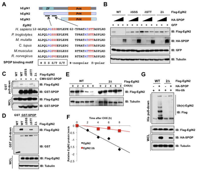
Fig. 4. SPOP interacts with and degrades EglN2 in a degron-dependent manner.
(A) A schematic illustration of the domain structures of EglNs and the alignment of binding motif of SPOP in EglN2 across different species. (B) IB analysis of WCLs derived from 293T cells transfected with the indicated constructs. (C) IB analysis of WCLs and GST pull-down products derived from 293T cells transfected with the indicated plasmid. Cells were treated with MG132 (10 μM) for 10 h before harvesting. (D) GST pull-down assay was performed with bacterially purified recombinant GST-SPOP proteins (GST as negative control) and the WCLs derived from 293T cells transfected with indicated EglN2 mutants. (E and F) IB analysis of WCLs derived from 293T cells infected with the indicated constructs. Where indicated, Cycloheximide (CHX) (100 μg/ml) was added after transfection 30 h, and the resulting cells were harvested at the indicated time points. Relative EglN2 protein abundance in (E) was quantified by Image J and plotted in (F). (G) IB analysis of WCLs and His pull-down products derived from 293T cells transfected with constructs encoding the indicated proteins and treated with MG132 (10 μM) for 10 h before harvesting.

