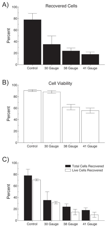
Figure 2.
Determination of minimal cannula size for safe passage of cells. Top panel: Percentage of recovered (both live and dead) cells after pipetting only (Control) and injecting RPE cells through 30, 38 and 41 gauge cannulas. Middle panel: Viability of the recovered cells measured as the proportion of live cells to total recovered cells. Viability remained constant between control and the 30g cannula (90%), but was reduced to ~50% in the 38 and 41g cannulas. Panel C combines panels A and B and illustrates the total cells recovered for each cannula and the cell viability within each recovered sample.

