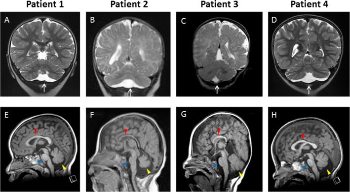
Figure 1.

Selected coronal and sagittal MR images in four patients. (A and E) Patient 1: A 3‐year‐old female with 2p15p16.1 deletion (3.24 Mb). The cranial MRI was performed at 2 years of age. (A) T2‐weighted coronal and (E) T1‐weighted sagittal MR images show (A) mild hypoplastic, flattened cerebellar hemispheres with proportionally reduced size of the vermis and dilated subarachnoid space beneath the cerebellum (white arrow), and (E) mild hypoplasia of the pons (blue arrowhead), a slightly small cerebellar vermis (yellow arrowhead), and intact corpus callosum (red arrowhead). (B and F) Patient 2: A 22‐month‐old female with 2p15p16.1 deletion (5.04 Mb). The cranial MRI was performed at 9 months of age. (B) T2‐weighted coronal and (F) T1‐weighted sagittal MR images show (B) hypoplastic, flattened cerebellar hemispheres with proportionally reduced size of the vermis with dilated subarachnoid space beneath the cerebellum (white arrow), and (F) prominent hypoplasia of the pons (blue arrowhead), very small cerebellar vermis (yellow arrowhead), and thin corpus callosum (red arrowhead). (C and G) Patient 3: A 4 years 2 months old male with 2p14p16.1 deletion (5.12 Mb). The cranial MRI was performed at 5 months of age. (C) T2‐weighted coronal and (G) T1‐weighted sagittal MR images show (G) normal size and shape of the cerebellar hemispheres, cerebellar vermis (yellow arrowhead), pons (blue arrowhead), and small corpus callosum (red arrowhead), and (C) without dilated subarachnoid space beneath the cerebellum (white arrow). (D and H) Patient 4: A 5‐year‐old male with 2p16.1 deletion (1.12 Mb). The cranial MRI was performed at 2 years of age. (D) T2‐weighted coronal and (H) T1‐weighted sagittal MR images show (D) mild hypoplastic, flattened cerebellar hemispheres with proportionally reduced size of the vermis with dilated subarachnoid space beneath the cerebellum (white arrow), and (H) mild hypoplasia of the pons (blue arrowhead), small cerebellar vermis (yellow arrowhead), and intact corpus callosum (red arrowhead).
